Photochromic FRET
The researchers of the Piston Lab have developed a technique utilizing optically-switchable fluorophores for high-sensitivity fluorescence resonance energy transfer (FRET) measurements. We can measure FRET efficiencies of <1% in live cells.
IMS-iSPIM
The system can be coupled to an image mapper spectrometer (IMS) to perform fast 3D spectral imaging.
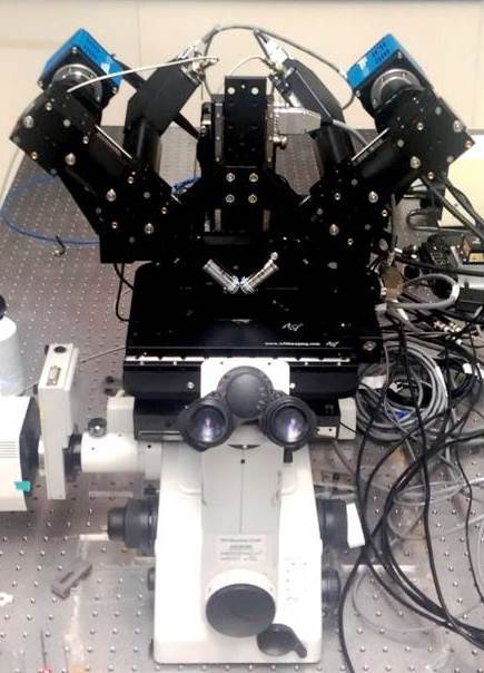
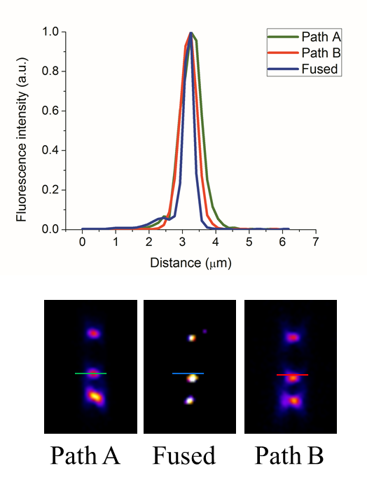
diSPIM
A dual-view inverted selective plane illumination microscope (iSPIM) is used to perform isotropic 3D fast imaging.
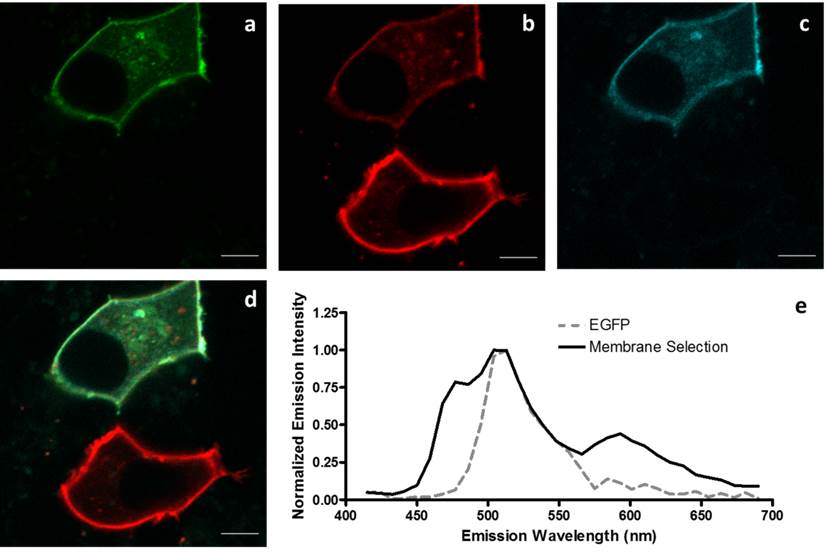
Fluorescent probes
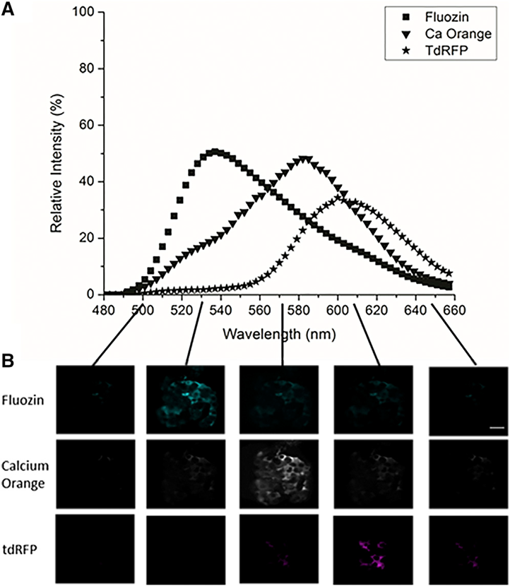
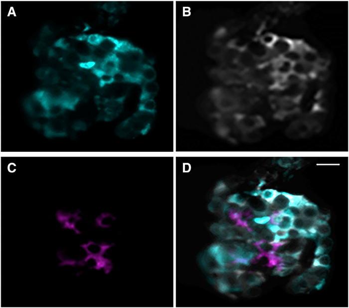
A library of VFP variants are maintained for multi-color imaging to simultaneously study protein function and localization.

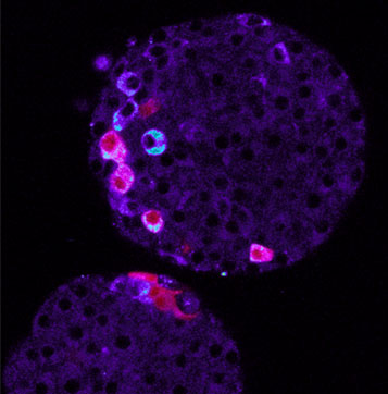
GPCR regulation of insulin secretion
Among other G-protein Coupled Receptors, dopamine receptors are interesting target of our research, for their effect on insulin secretion and calcium oscillations.
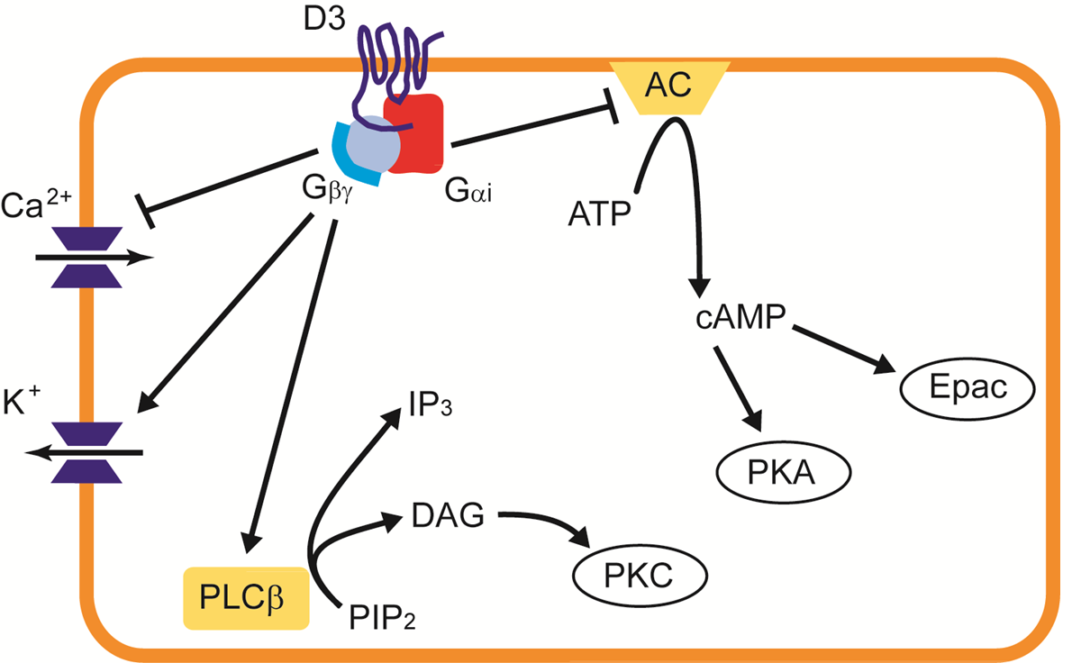
We tag the GPCR, the Gβγ complex and its possible targets to study intra-cellular signaling in live cells.
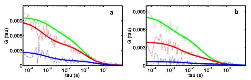
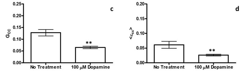
FCCS, PCH and SpIDA analysis techniques are used to study these protein-protein interactions in live cells.
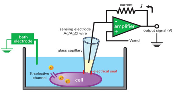
Membrane potential
Using whole cell patch clamp techniques (top), we can measure the channel gating of Ca2+, Na+, and K+ channels associated with hormone secretion. We also use dyes such as DiSBAC to measure plasma membrane potential with fluorescence microscopy (bottom).
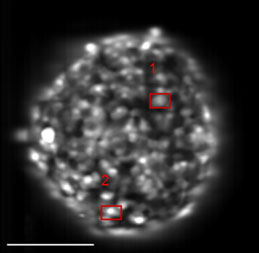
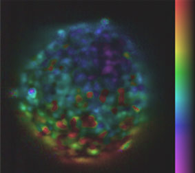
Metabolic state imaging
NAD(P)H autofluorescence reports cellular glucose metabolism. Metabolic dynamics are measured across the islet in response to increasing concentrations of glucose.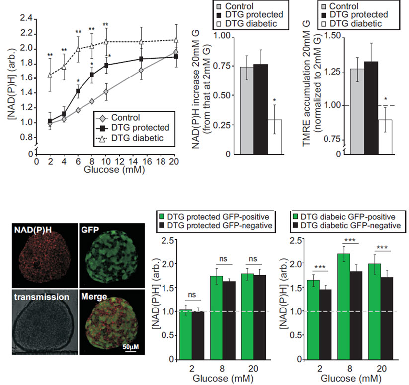
Microfluidics
We create and utilize novel microfluidic devices to isolate pancreatic islets for live imaging under a variety of conditions.
Islet Hemodynamics
We are developing a semi-automated method to monitor and analyze in detail the in vivo blood flow in the pancreas of living mice using confocal microscopy, fluorescent labels, and extensive computer software.
Ca2+ Oscillations
At elevated glucose levels, synchronized oscillations in intra-cellular free calcium ([Ca2+]i) are observed. High-speed imaging shows calcium waves propagate across the islet with a velocity of ~80μm/s.
Without gap junction coupling, beta cells show unsynchronized [Ca2+]i oscillations (left). As we reduce gap junction coupling, our quantitative model explains the loss in synchronization and reduction in wave velocity (right).
Brown Fat (BAT)

Subcutaneous brown adipose tissue (BAT) transplants in mouse models of type 1 diabetes result in euglycemia, weight gain, and reversal of clinical signs of diabetes.
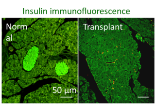
Importantly, the effects of BAT transplants are independent of insulin. We are currently exploring the molecular mechanisms behind this physiological observation.
Gunawardana S. and Piston DW , Diabetes (2012)